MOLECULAR ARCHITECTURE OF VIRUSES
Research Interests
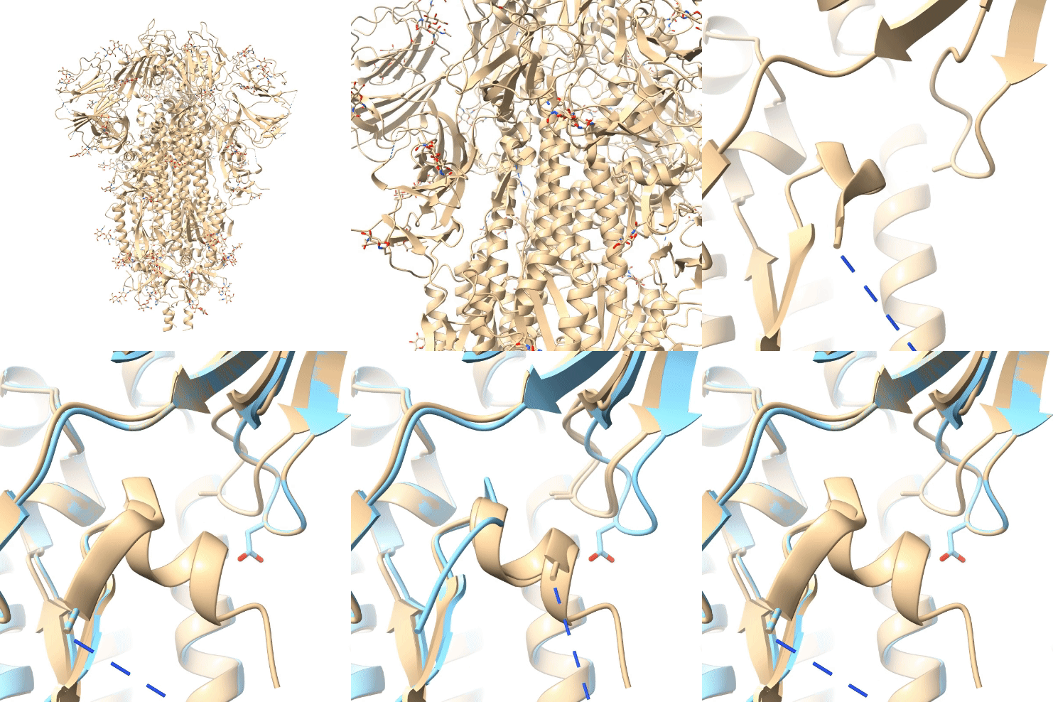
Research in the Ke Lab aims to understand viral structure and function by dissecting key macromolecular complexes upon host-pathogen interactions. We will take advantage of the state-of-the-art technique of cryo-electron microscopy (cryo-EM) and cryo-electron tomography (cryo-ET), which allows high-resolution structure determination of macromolecules in their native environment. We are also interested in developing and applying machine learning to process low signal-to-noise ratio micrographs. Ultimately, the Ke Lab aims to uncover molecular insights into cancer-causing viral diseases, providing a basis for antiviral therapy and vaccine development.
Below are examples of cryo-ET applications in investigating viruses and their interactions with host cells.
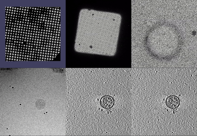
Journey to find a SARS-CoV-2 virion: cryo-ET
Zoom-in view from a TEM grid (3 mm in diameter) into a square (~100 µm), then into a hole (2 µm in diameter), then into a single SARS-CoV-2 virus (~100 nm in diameter).
Images were taken from a Titan Krios transmission electron microscope operating at cryogenic temperature (around -190 ºC).
A series of images at different tilt angles are presented, followed by a 3D tomographic reconstruction of the virion. Spike trimers protruding away from the viral membrane are visible.
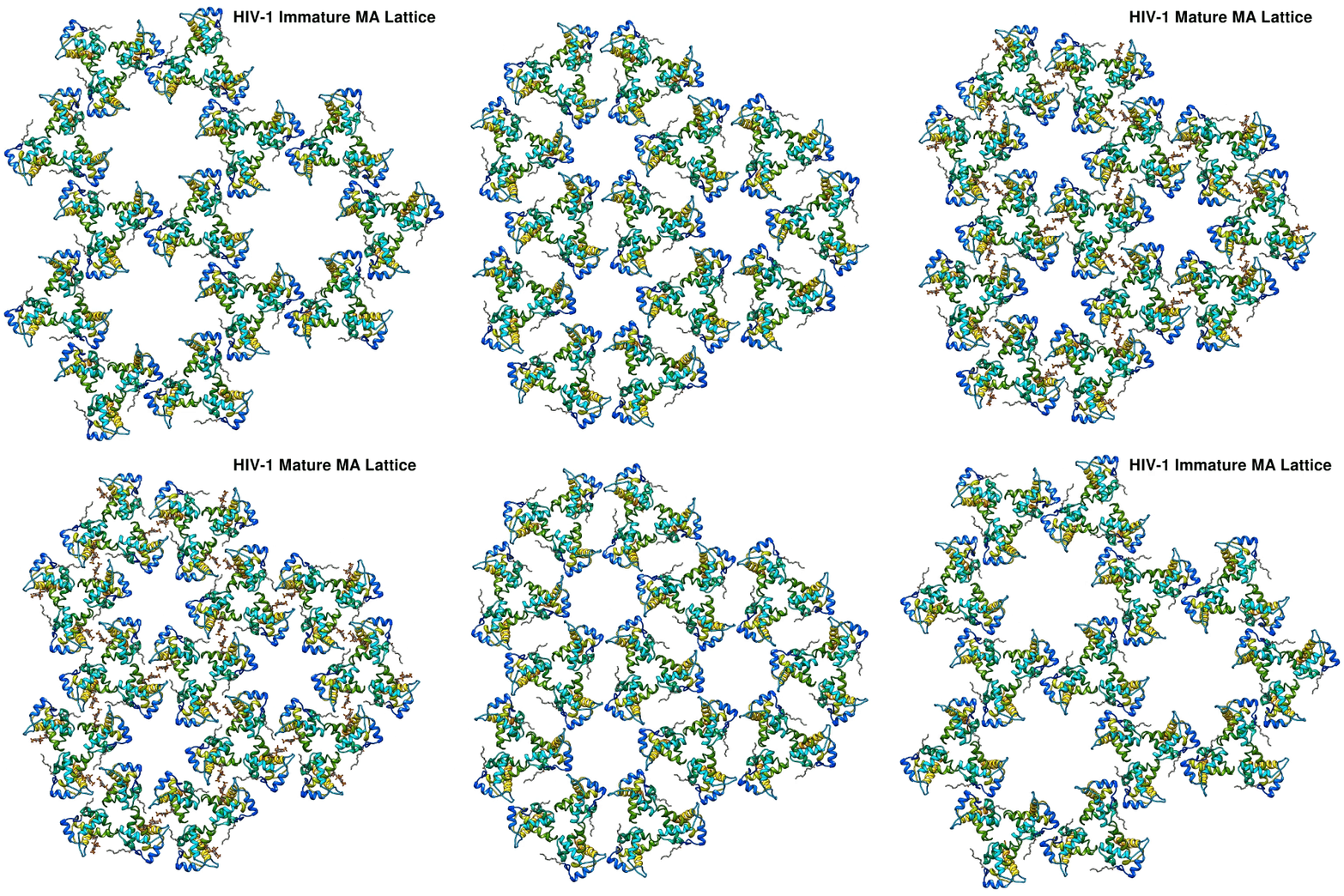
HIV-1 maturation
HIV-1 Matrix protein undergoes substantial structural maturation to form very different lattices in immature and mature HIV-1. This is achieved by investigating mature and immature HIV-1 virions, followed by subtomogram averaging to high-resolution (sub-nanometer) to resolve the structural differences in these two different lattices.
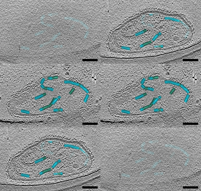
Respiratory Syncytial Virus Assembly
Respiratory syncytial virus assembly stages are captured by cryo-electron tomography from virus-infected cells. Here, initiation, elongation, assembly, and scission events are captured in one single micrograph. Green is the membrane, red is the fusion glycoprotein, and magenta is the viral ribonucleoprotein complex (RNP).


